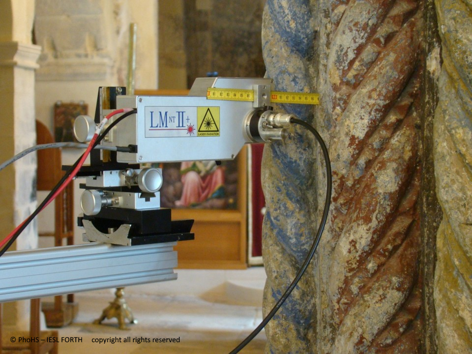
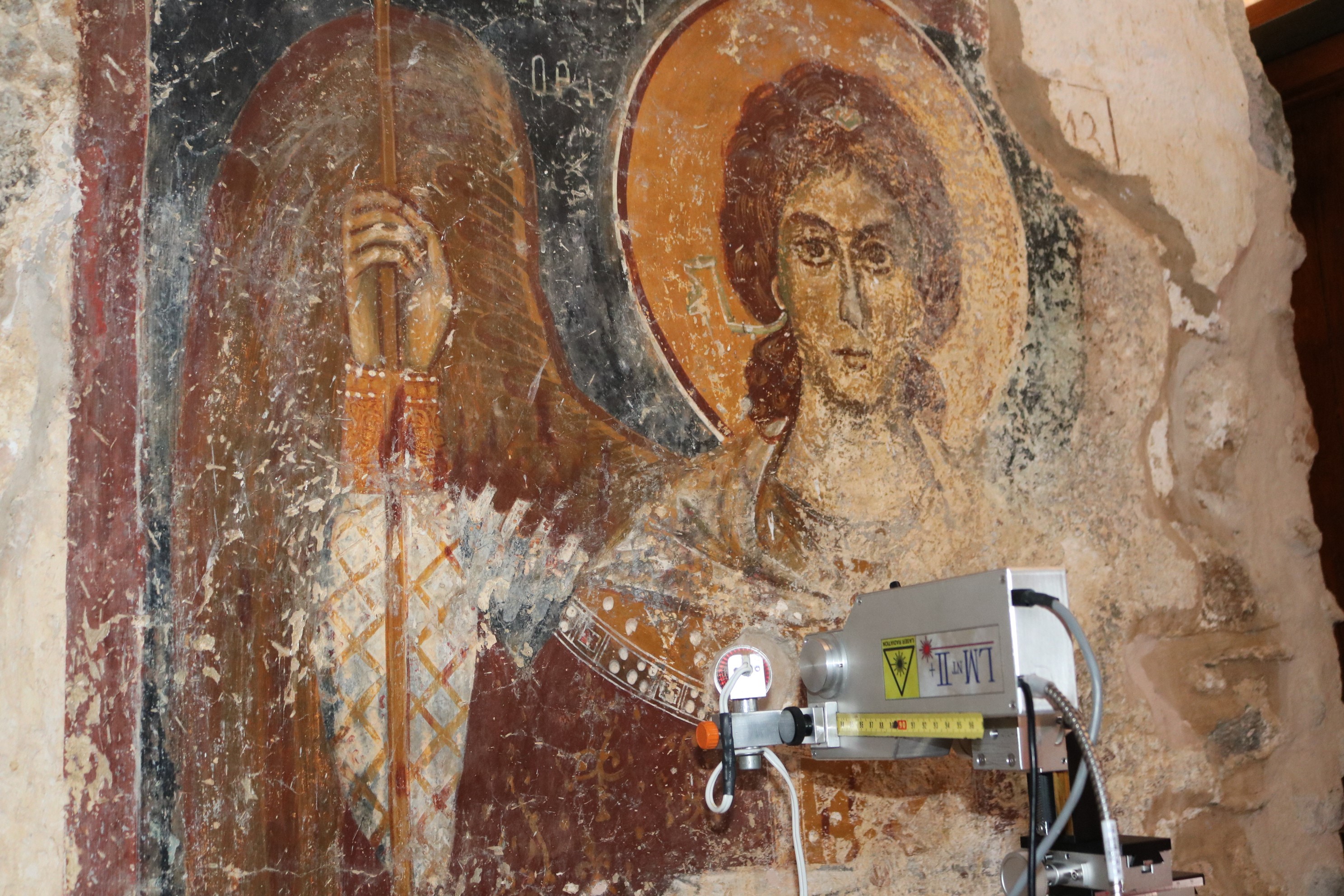
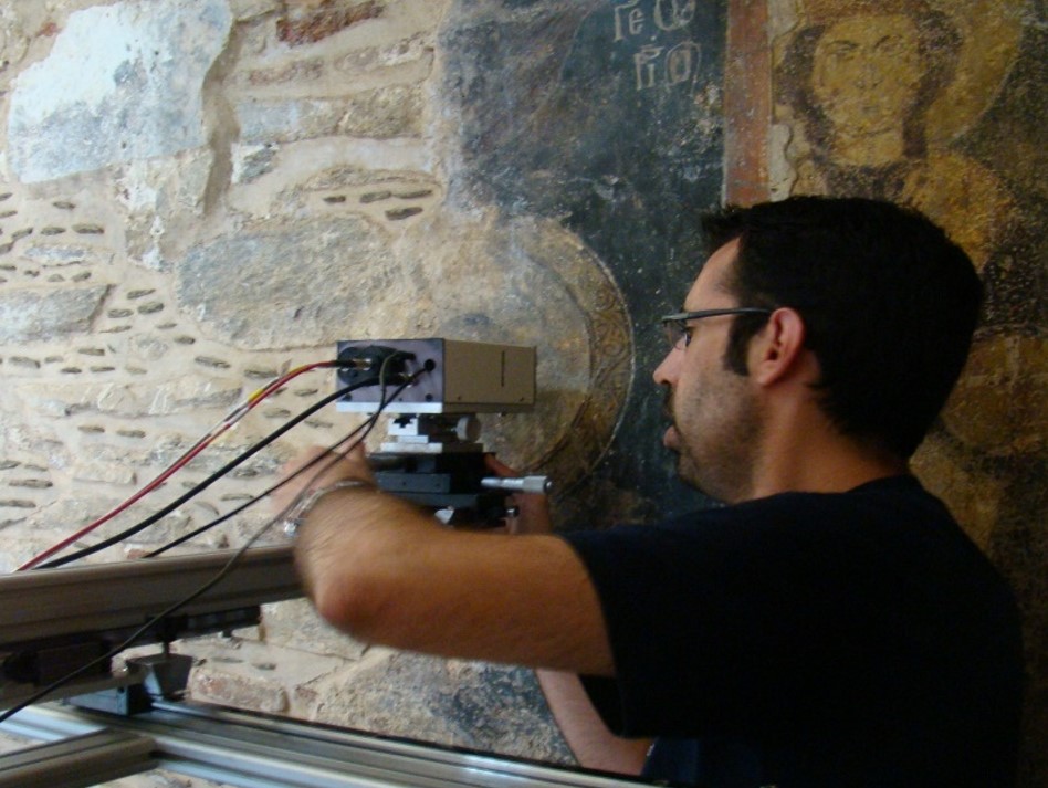
E-RIHS.GR THE GREEK NODE OF THE EUROPEAN RESEARCH INFRASTRUCTURE FOR HERITAGE SCIENCE
Start Date: 01/02/2017,     End Date: 30/09/2020

E-RIHS.gr is the Greek node of the European Research Infrastructure for Heritage Science, E-RIHS.
E-RIHS.gr offers

  1.  interdisciplinary access to novel scientific facilities and technologies, though standardized techniques and protocols, for the study, the documentation and the protection of Cultural  Heritage, and 
  2. to scientists and professionals related to Heritage science, advanced training on innovative methods for the analysis, diagnosis and conservation of CH objects and monuments.


E-RIHS.gr is the Greek cross-disciplinary Research Infrastructure (RI) in the service of Heritage Science (HS).

E-RIHS.gr exploits the expertise sourcing from the long term involvement of our partners in the field of research for HS, the know-how derived from the participation in numerous EU and National projects related to HS, as well as the leading infrastructures and state of the art instruments available in our laboratories, in order to deliver Innovative, Reliable and Efficient tools for addressing demanding  challenges in Cultural Heritage research.

Our concept is described in three words/terms:

Micro methods and tools appropriate for analytical studies of CH objects and materials, namely at the microscopic level.

These involve novel laser/spectroscopic techniques for compositional analysis and mapping of materials on works of art, advanced methods for studying archaeological DNA and also modern laser-based technologies for conservation and restoration.

Macro: Non-invasive geophysical and geochemical measurements for surveying and mapping macroscopically the landscape of archaeological and historical sites including underwater ones.

Info: Development and use of innovative management systems that enable efficient documentation and handling of new data and information enhancing our understanding about CH sites, monuments and objects.


-RIHS.gr, under the name IPERION-CH.gr, also participates in the Hellas-CH facility which is included in the Greek National Roadmap for Research Infrastructures.

E-RIHS will provide access to a wide range of cutting-edge scientific infrastructure, methodologies, data and tools, training in the use of these tools, public engagement, access to repositories for standardized data storage, analysis and interpretation. E-RIHS will enable the community to advance heritage science and global access to the distributed infrastructures in a coordinated and streamlined way.

E-RIHS.gr provides access services through three integrated platforms: 

 MOLAB  Mobile instruments for in-situ diagnostics - The MObile LABoratory allows  users to access state of the art mobile instrumentation for the study and diagnosis of valuable or immovable objects, archaeological sites and historical monuments.

FIXLAB  Access to advanced laboratories - The FIXed LABoratory provides access to leading infrastructures for sophisticated scientific investigations on samples or whole objects

DIGILAB Digital documentation &  management - The DIGItal LABoratory allows users  to exploit advanced databases for the documentation of their research and the handling of their data.


If you are interested to apply for E-RIHS access -  services

browse the E-rihs.gr application form :https://www.e-rihs.gr/contact/

Prof. Anglos Demetrios
University Faculty Member
Dr. Pouli Paraskevi
Senior application Scientist
Prof. Sotiropoulou Sophia
University Faculty Member

The hybrid mobile system combines three spectroscopic techniques: Laser-Induced Breakdown Spectroscopy (LIBS), Diffuse Reflectance Spectroscopy (DR) and LED-Induced Fluorescence (LED-IF), leading to an integrated characterization of the material under examination ‘in-situ’ with no sample removal or preparation restrictions. Moreover, the rapid data acquisition and the user-friendly software increase the applicability of the system in archaeometry and art conservation.

LIBS offers qualitative and semi-quantitative multi-elemental analysis as well as the ability to perform stratigraphic analysis on a multi-layered object. A compact Nd:YAG laser (1064 nm, τΡ = 10 ns) is focused by means of a lens on the surface of the object generating a micro-plasma, whose emission is transmitted via a bifurcated optical fiber into a dual spectrometer unit (Avaspec-2048-2-USB2, Avantes) (spectral range: 200 - 660 nm, resolution: 0.2 - 0.3 nm) and the analytical information is acquired within seconds. 

DR and LED-IF techniques reveal information about the molecular composition of a material; DR is based on the absorption of light from the material and LED-IF is based on the fluorescence induced from a LED source. Both techniques are non-destructive, making them extremely attractive for the analysis of objects in cultural heritage and archaeology. A halogen tungsten lamp is used for the DR measurements, while for the LED-IF measurements a LED source (375, 438 or 632 nm) excites the material. In order to achieve a broader spectral coverage, the signal from the DR and LED-IF is recorded via a low resolution spectrometer (Avaspec-2048L-USB2, Avantes) (spectral range: 200 - 1100 nm, resolution ~ 2.5 nm).

A miniature CCD camera offers a magnified view of the object surface during analysis and permits accurate aiming of the laser beam with the aid of a cross-hair indicator superimposed on the image.

All the light sources along with the necessary optics and visualization camera are integrated in a lightweight and compact optical probe head.


The LIBS microscope has the capacity to provide fast elemental mapping of flat surfaces, typically cross-sections of geological samples, marine shells, bones, teeth etc. The 2D-elemental maps of the scanned surface can be used to identify the distribution of mineral phases in rocks, to measure the variability of elemental proxies related to paleoenvironment in shell studies or to assess the diffusion of environmental pollutants into hard tissues. 

In the present micro-LIBS workstation a Q-switched Nd:YAG laser is used (λ = 1064 nm, pulse duration: 10 ns, pulse energy 5-20 mJ). The laser beam is focused on the sample through a laser objective lens down to a spot of 40 - 60 μm in diameter. The light emitted by the plasma is transferred via an optical fiber to the spectrometer which captures LIBS spectra for each one of the laser pulses that scans the surface. According to specific analysis requirements the spectral data is processed on-line or following completion of scanning. Samples are mounted on a motorized X–Y–Z micrometric stage and translated with respect to the laser beam that remains in a fixed position. The typical translation step is of the order of 100 μm. A CCD camera enables the user to have a clear view of the sample surface and to define the area that is to be mapped (any shape is acceptable). The measurement speed is about 0.9 s per point and each map could have 500 - 6000 points (i.e. an elemental map of 4000 points is obtained in about 1 hr). A dual spectrometer unit (AvaSpec-2048-2-USB2) records the LIBS signal (spectral range: 200 - 640 nm, resolution ~ 0.2 - 0.3 nm). For higher sensitivity, the signal can be recorded by a Czerny-Turner spectrometer (Jobin Yvon, TRIAX 320) with an ICCD camera (DH734–18F, Andor Technology) (spectral range ~ 45 nm).

Raman spectroscopy gives details about the molecular structure of samples on the basis of the characteristic vibrational modes of the molecule. It can be used to identify a variety of materials (minerals, pigments, organics etc.) and the fact that it is completely non-destructive makes it extremely attractive for the analysis of invaluable objects, such as artworks and archaeological objects.

Our mobile Raman microspectrometer (Exemplar Plus, ΒWTEK) uses a cw (continuous wave) diode laser working at 785 nm as excitation source. An optical probe head focusses the laser beam onto the sample surface by means of a set of objective lenses offering different levels of magnification. A white LED and a digital colour camera are included on the optical head, which allow visualization of the object’s surface and selection of the area (spot) to be analyzed. The spectrometer provides high spectral resolution (< 8 cm−1) and sensitivity, covering a spectral range between 100 – 3300 cm−1. The employed detector (Peltier-cooled) features high sensitivity with low dark counts.


IRIS-II, a portable MultiSpectral Imaging instrument, enables the compositional and structural study of multi-layered Cultural Heritage surfaces. It is fully portable enabling thus the examination of objects in situ (museums, conservation laboratories, archaeological areas etc.). This imaging system, provides detailed information related to the physical and chemical properties of materials, based on reflection and fluorescence spectroscopy.

The main elements of the multi-spectral imaging system include a camera, an imaging monochromator equipped with a filter wheel of 28 band-pass filters, the objective lens, electronics and a computer that controls all the components. The camera used on the system is a monochrome digital CMOS camera. The spatial resolution is 5MPixels, while the dynamic range applied is 8 bit. This sensor is sensitive from 350 nm up to 1200 nm. 

The whole system is designed to be portable and can be carried in a small case. 

Finally, custom-made software, entirely developed in LabView is employed. This software enables the control of the system and the data acquisition. Additional processing software for images normalization, calibration and analysis is also developed and used.

IESL-FORTH holds a number of laser systems with different wavelength, pulse duration and energy output characteristics available for laser cleaning investigations such as:

  • Transportable Q-switched Nd:YAG lasers (Quantel Q-smart 850, LITRON TRLi, Spectron SL-805 modified, Quanta Palladio, BMI 5022 DNS 10) emitting both nano- and pico-second (EKSPLA SL 312) laser pulses at various wavelengths (such as 1064, 532, 355, 266 & 213 nm)
  • Various excimer lasers emitting nano, pico and femto-second pulses in the UV
  • A patented transportable ns Nd:YAG system with dual-wavelength beam output, developed for the laser cleaning project of the Athens Acropolis Monuments especially dedicated to remove pollution crust from stonework without any discoloration or damage
  • A transportable LQS Nd:YAG system (ElEn, EOS1000) emitting IR pulses at longer pulse-widths
  • An Er:YAG laser system (LITRON NANO L 200-20-Er) emitting at 2094 nm 
  • A continuous CO2 laser system (Coherent Diamond C20) for the patented application related to the laser conservation of glazed objects.

Various workstations adaptable for different laser cleaning applications with the ability to integrate different optical and opto-mechanical components for the most appropriate beam delivery and control are available such as:

  • Handheld units (using a articulated mirrored arm) 
  • Automated beam scanning units for micrometer control and guidance of the laser beam to the sample (i.e. the painting surface).

The latter, a computer-driven mechanized component, can be adjusted on the basis of fluence values, spot size and pulse repetition rate enabling thus the homogeneous scanning of predefined areas.

Furthermore, a number of multi-modal diagnostic instruments for in-situ assessment of the cleaning result and monitoring of the laser ablation procedure are also availableThese can be selected according to the specifications of each individual cleaning case and may be one or more of the following:

  • Spectral Imaging to visualise the cleaning state
  • Laser-Induced Fluorescence (LIF) to evaluate the thinning of varnish
  • Vis-NIR Diffuse Reflectance spectroscopy to chemically characterise the irradiated surfaces

The DHSPI (Digital Holographic Speckle Pattern Interferometry) systems have been developed and continuously optimized at IESL-FORTH with the aim to investigate and monitor deformation, deterioration, and fracture mechanisms and thus to evaluate the structural condition of materials and systems as a result of ageing, mechanical alteration and materials’ failure.
DHSPI captures microscopic alterations of sub-surface topography on the basis of high-resolution interferometric imaging. Hidden defects are revealed as visible interference fringe patterns forming locally inhomogeneous intensity distribution patterns. The deformation data are extracted through the differential displacement of the surface under investigation and the deformation value is measured by multiples of half wavelength.

DHSPI-II, the most recent model, is a compact fully portable system with a built-in data acquisition and processing unit and dedicated user-friendly software, for the system control and data post-processing which enables real-time qualitative and quantitative structural diagnosis. DHSPI-II also allows control (via cable) from a remote pc (eg laptop) which provides extra flexibility for in-situ measurements. 

TECHNICAL INFO:  •Laser power: 300mW     •Coherence length: >30m     •CCD resolution: 5MP    •Spatial resolution: 144 lines/mm     •Displacement resolution: ≥ 266nm    •Sensor lens: C-Mount type exchangeable   •Beam Divergence: >40cm@1m (Gaussian Profile)